67 results filtered with: Photograph

- Digital Images
- Online
X-ray of a knee joint printed on coated aluminium, artwork
Chiara Salvi
- Digital Images
- Online
Exhibition '100 Years of medical Photography', 1955.

- Digital Images
- Online
Kidneys showing bilateral atrophy (tissue wasting)
Michael Frank, Royal Veterinary College
- Digital Images
- Online
A man scything. Photogravure after Eadweard Muybridge, 1887.
Eadweard Muybridge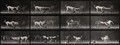
- Digital Images
- Online
Three dogs fighting over a piece of cloth. Photogravure after Eadweard Muybridge, 1887.
Eadweard Muybridge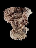
- Digital Images
- Online
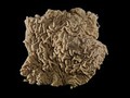
Cow foregut showing multiple warty growths (papillomas). These have grown from the gut lining, which is formed of squamous epithelium (consisting of flat, thin cells). These benign (non-cancerous) tumours can be caused by papillomavirus infection.
Michael Frank, Royal Veterinary College
- Digital Images
- Online
Tropical Diseases, Elephantiasis of legs.

- Digital Images
- Online
E. Muybridge "Animal locomotion", plate

- Digital Images
- Online
X-ray of a knee joint printed on coated aluminium, artwork
Chiara Salvi
- Digital Images
- Online
E. Muybridge "Animal locomotion", plate

- Digital Images
- Online
Dog (puppy) with cleft palate
Michael Frank, Royal Veterinary College
- Digital Images
- Online
Tropical Diseases, Elephantiasis of the scrotum.
- Digital Images
- Online
Horse intestine with multiple attached parasitic worms
Michael Frank, Royal Veterinary College
- Digital Images
- Online
A woman putting a barrel on her shoulder. Photogravure after Eadweard Muybridge, 1887.
Eadweard Muybridge
- Digital Images
- Online
A woman bearing basket on head. Photogravure after Eadweard Muybridge, 1887.
Eadweard Muybridge
- Digital Images
- Online
Canine skull with osteosarcoma
Michael Frank, Royal Veterinary College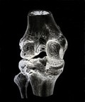
- Digital Images
- Online
X-ray of a knee joint printed on coated aluminium, artwork
Chiara Salvi